Dental X-rays – Are They Safe and Necessary?
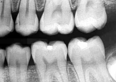
“A picture is worth a thousand words” and there is no exception to this for your dentist. Dental radiographs (images created with the use of ionizing radiation) are dental pictures that serve as an important tool to aid the human eye of the dentist in the detection and diagnosis of diseases, lesions, and health of the teeth and bone. Radiographs serve as an extra pair of eyes for seeing things that can’t be seen by just looking in the mouth.
The dentist should determine the patient’s need for radiographs based on the individual patient’s medical history, intraoral dental examination, oral health history, risks and benefits, and previous radiation exposure to medical and/or dental x-radiation. Since 1987, the American Dental Association (ADA), along with the U.S. Food and Drug Administration (FDA), has adopted guidelines for prescribing dental radiographs to assist the dentist in deciding how often and what type of radiographs to take.
Once the need for dental radiographs has been decided, trained dental professionals are expected to use radiation safely. Dental offices use the ALARA (as low as reasonably achievable) principle that states all exposures to radiation must be kept to a minimum. This is done in several ways such as the use of digital sensors which can reduce the dose from 50 to 90% compared to traditional film, a lead apron with a thyroid collar, sensor holders to stabilize the sensor in the mouth, appropriate exposure time and settings, and proper technique to ensure the quality of the image.
Radiation is naturally present in our environment. People are exposed to ionizing radiation in the natural environment (known as background radiation) on a daily basis. Natural environmental sources may include cosmic rays from outer space e.g., flying in an airplane, radioactive elements in some foods like bananas, the ground (terrestrial) and radon gas (a gas from the earth’s crust), to name a few. According to the U.S. Nuclear Regulatory Commission (U.S.NRC), “about half the total annual average U.S. individual’s radiation exposure comes from natural sources. The other half is mostly from diagnostic medical procedures.”
The amount of ionizing radiation used in dentistry is considered a low dose because it is focused just on the mouth. For example, a full mouth series of digital radiographs is equal to approximately 20 days of exposure to background radiation. The risk of getting cancer from dental x-rays would be comparable to a 1 in 1 million risk of death due to an accident while traveling 1000 miles in airplane or 300 miles in an automobile. One must also keep in mind that the human body is composed of trillions of cells. Cells have ways of repairing damage that may be caused by radiation.
Similar to digital cameras, digital radiography has simplified and improved the process of using dental radiation and reduced the risks. Ask your dental professional to explain the risks, benefits, and precautions they take to keep you safe.
About the Author:

Kaye Raasch, RDH, CDA, MS has more than 35 years’ experience in dental profession. She currently is a dental hygiene educator in the dental hygiene program at Northeast Wisconsin Technical College in Green Bay, Wisconsin where she has devoted 25 years to her passion of teaching. She enjoys planting seeds of knowledge in her students and watching them grow. Her teaching expertise is in dental radiography, clinical dental hygiene, special needs and oral histology/embryology. She has volunteered in several Mission of Mercy dental outreach venues in the Wisconsin area. She is a member of the American Dental Hygienists’ Association and WI Dental Hygiene Association.
Offers From Our Partners: